
Essential Reconstruction for Speech and Function
Cleft palate—a gap in the roof of the mouth—is a congenital condition requiring surgical repair to enable normal speech development, safe feeding, and proper ear function. Unlike cleft lip which is primarily visible, cleft palate profoundly affects function, making timely surgical correction essential. Modern cleft palate surgery achieves excellent outcomes, with most children developing normal or near-normal speech when surgery is performed at the appropriate age by experienced specialists within a comprehensive multidisciplinary care framework.
- Functional Priority:
Essential for normal speech development, safe swallowing, and middle ear ventilation - Critical Timing:
Typically performed at 9-12 months to optimize speech development before language explosion - Complex Reconstruction:
Requires meticulous repair of multiple tissue layers creating functional palatal mechanism - Speech Outcomes:
Most children achieve normal or near-normal speech with timely surgery and speech therapy - Lifelong Monitoring:
Requires ongoing multidisciplinary care through childhood into adolescence for optimal outcomes
Understanding Cleft Palate
Cleft palate occurs when the tissues forming the roof of the mouth fail to join completely during early pregnancy (weeks 6-9 of development). This creates a gap or opening connecting the mouth and nasal cavity. Cleft palate may occur alone or in combination with cleft lip, and varies considerably in severity.
Types of Cleft Palate
Soft Palate Only: The cleft affects only the soft palate (the mobile muscular portion at the back of the mouth) whilst the hard palate (bony front portion) is intact. This is the mildest form.
Soft and Hard Palate: The cleft extends through both soft and hard palate, creating complete communication between mouth and nose. This is the most common presentation requiring surgical repair.
Submucous Cleft: The surface mucosa (lining) is intact, but underlying muscle is cleft. Signs include bifid uvula (split), midline furrow in soft palate, or notch in hard palate. Submucous clefts may cause speech problems despite normal appearance.
Unilateral vs. Bilateral: The cleft may affect one side (unilateral) or both sides (bilateral) of the palate, with bilateral clefts being more severe.
Associated Features
Cleft palate affects the levator veli palatini muscles (responsible for closing the palate against the back of throat during speech and swallowing), which are abnormally positioned and must be repositioned during surgery. The Eustachian tubes (connecting middle ear to throat) function poorly, causing middle ear fluid and hearing problems. The maxilla (upper jaw) may be affected with dental and orthodontic implications.
The Functional Impact of Cleft Palate
Speech Development
The palate is essential for normal speech. During speaking, the soft palate elevates and closes against the back of the throat (velopharyngeal closure), directing air through the mouth for most sounds. With cleft palate, this closure is impossible; air escapes through the nose causing hypernasal speech (excessive nasal resonance) and difficulty producing certain sounds, particularly pressure consonants (p, b, t, d, k, g). Without repair, normal speech cannot develop. Timely surgery followed by speech therapy is essential for optimal speech outcomes.
Feeding Difficulties
Infants with cleft palate cannot create adequate suction for normal breastfeeding or bottle feeding, as the gap prevents building pressure in the mouth. Feeding takes longer and is less efficient. Milk may enter the nasal cavity causing choking or nasal regurgitation. Special feeding bottles with modified teats help manage feeding before surgery, but repair ultimately resolves these difficulties.
Ear Problems
The abnormal palatal muscles affect Eustachian tube function, preventing proper middle ear ventilation. This causes persistent middle ear fluid (glue ear) leading to conductive hearing loss. Chronic fluid increases infection risk. Most children with cleft palate require grommets (ventilation tubes) to maintain hearing during critical language development years. Palate repair improves Eustachian tube function, though ear problems may persist.
Dental and Orthodontic Issues
Cleft palate often involves the alveolus (gum ridge), creating gaps in the dental arch. This affects tooth eruption, position, and number (missing or extra teeth common). Orthodontic treatment and sometimes alveolar bone grafting are required to optimize dental alignment.
Cleft Palate Repair Surgery
Timing of Surgery
Cleft palate repair is typically performed at 9-12 months of age. This timing balances several factors: early enough to optimize speech development (before the language explosion around 12-18 months), late enough that the infant is larger and stronger for safer anaesthesia, and allowing time for the palate to grow, facilitating easier repair. Some surgeons prefer earlier repair (6-9 months) whilst others wait until 12-15 months; individual cleft team protocols vary.
Surgical Goals
Palate repair aims to close the cleft completely, separating oral and nasal cavities, reconstruct the levator veli palatini muscle sling (repositioning muscles into correct anatomical position for velopharyngeal function), create adequate palatal length for speech, preserve growth potential of the maxilla (minimizing surgical interference with facial growth), and establish foundation for normal speech development.
Surgical Techniques
Multiple surgical techniques exist; selection depends on cleft type, width, and surgeon preference. Common approaches include:
Two-Flap Palatoplasty: Mucoperiosteal flaps are raised from both sides of the cleft and brought together in the midline, closing the hard palate. The soft palate muscles are dissected, repositioned, and sutured to reconstruct the levator sling.
Intravelar Veloplasty: Specific attention to radical dissection and repositioning of the levator muscles, creating a functional muscular sling in the soft palate essential for speech.
Furlow Double-Opposing Z-Plasty: Uses geometric Z-shaped incisions to lengthen the soft palate and reposition muscles simultaneously. This technique is particularly effective for soft palate clefts.
Vomer Flaps: For wide clefts, tissue from the nasal septum (vomer bone) is used to help close the nasal layer.
The Procedure
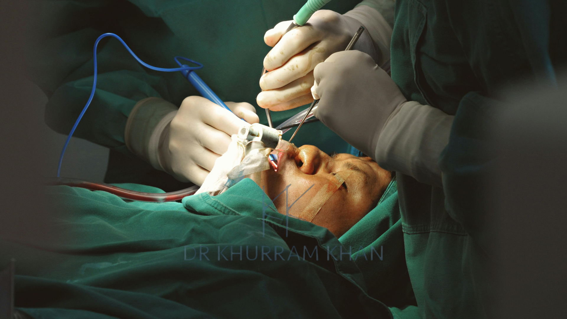
Surgery is performed under general anaesthesia, taking 2-3 hours. The mouth is held open with a specialized retractor. Using magnification, the surgeon meticulously dissects tissues, separates oral and nasal layers, repositions muscles, and closes all layers carefully. Multiple rows of dissolving sutures close the repair. No external incisions are made; all work is intraoral.
Recovery and Outcomes After Palate Repair
Immediate Post-Operative Care
Infants typically stay in hospital 1-2 nights. Pain management is crucial using regular analgesia. Feeding resumes carefully with soft foods or liquid diet for 2-3 weeks, avoiding hard items that could damage the repair. Arm splints may be used temporarily preventing the infant from putting fingers in the mouth. The palate appears swollen initially with dissolving sutures visible. Healing progresses rapidly over 2-3 weeks.
Speech Development
The primary goal of palate repair is enabling normal speech. Most children (70-80%) develop normal or near-normal speech following timely repair and appropriate speech therapy. Early speech therapy assessment (around 18 months) identifies any difficulties, with therapy commencing if needed. Some children develop velopharyngeal insufficiency (VPI)—inadequate closure of the soft palate causing persistent hypernasal speech—requiring further intervention.
Hearing and Ear Care
Grommets are often inserted during palate repair to drain middle ear fluid and maintain hearing. Regular audiology monitoring throughout childhood ensures hearing remains adequate for language and learning. Some children require multiple sets of grommets until Eustachian tube function improves.
Long-Term Outcomes
With modern techniques and comprehensive care, outcomes are excellent. Most children achieve intelligible speech, attend mainstream education, lead normal lives, and require minimal ongoing intervention beyond orthodontic care. However, this depends critically on timely surgery, consistent speech therapy, regular monitoring, and appropriate secondary procedures when needed.
Secondary Procedures
Speech Surgery (Velopharyngeal Surgery)
If VPI persists despite speech therapy, secondary speech surgery may be necessary. Options include pharyngeal flap (tissue from the back of throat creates a bridge to the soft palate, reducing nasal air escape), sphincter pharyngoplasty (repositioning pharyngeal muscles to narrow the velopharyngeal port), or palate re-repair. These procedures typically occur around age 4-7 once speech patterns are established and the problem clearly defined.
Alveolar Bone Grafting
For clefts involving the alveolus (gum ridge), bone grafting—typically around age 8-10 before permanent canine tooth eruption—fills the bony gap, provides support for teeth, and stabilizes the dental arch. Bone is usually harvested from the hip.
Orthognathic Surgery
Some patients develop maxillary hypoplasia (underdeveloped upper jaw) causing malocclusion (bite problems) and facial imbalance. Orthognathic surgery in adolescence (16+) advances the upper jaw, improving bite and facial aesthetics.
Palatal Fistula Repair
Small holes (fistulae) may develop in the palate repair, causing liquid escape into the nose or hypernasal speech. Fistula repair, often challenging, closes these openings.
The Essential Role of Specialist Multidisciplinary Care
Cleft palate management requires coordinated multidisciplinary care from birth through adolescence. In the UK, care is centralized in specialist cleft centres ensuring expertise and optimal outcomes.
The Cleft Team
Comprehensive care involves cleft surgeons (plastic surgeons and oral surgeons), speech and language therapists, orthodontists, ENT surgeons, audiologists, paediatricians, geneticists, psychologists, and specialist cleft nurses. This team provides coordinated assessment, treatment planning, and lifelong monitoring.
Specialist Surgical Expertise
Mr Khurram Khan is a distinguished Consultant Plastic Surgeon with advanced fellowship training specifically in cleft lip and palate surgery at Birmingham Children’s Hospital, a leading UK cleft centre. His expertise encompasses primary palate repair in infants, complex secondary speech surgery, palatal fistula repair, and comprehensive cleft care within a multidisciplinary framework.
His commitment to achieving optimal functional and aesthetic outcomes, combined with understanding of growth, speech development, and family needs, ensures patients receive exceptional care throughout their cleft journey.
Accessing Care
Cleft palate surgery is provided through the NHS at specialist cleft centres. Families are referred during pregnancy or shortly after birth, with coordinated care continuing through childhood. For patients seeking specialist assessment or second opinions, contacting Mr Khan’s practice provides access to expert evaluation and guidance.
Frequently Asked Questions About Cleft Palate Surgery
At what age is cleft palate surgery performed?
Cleft palate repair is typically performed at 9-12 months of age. This timing optimizes speech development by repairing before the language explosion (12-18 months) whilst allowing sufficient infant growth for safer anaesthesia. Some centres prefer slightly earlier (6-9 months) or later (12-15 months) repair; your cleft team will recommend optimal timing based on individual circumstances and their protocols. If cleft lip is also present, it is repaired first at approximately 3 months, followed by palate repair several months later.
Will my child’s speech be normal after cleft palate repair?
Most children (70-80%) develop normal or near-normal speech following timely palate repair and appropriate speech therapy. Speech outcomes depend on timing of surgery (earlier repair generally better), quality of surgical repair, consistent speech therapy when needed, and individual factors. Some children develop velopharyngeal insufficiency causing persistent hypernasal speech, requiring further intervention. Early speech assessment and therapy significantly improve outcomes. Realistic expectations are important; whilst most children achieve excellent speech, some require additional procedures or ongoing therapy. Your cleft team monitors speech development closely throughout childhood.
Will my child need more than one palate operation?
Most children require only the primary palate repair. However, approximately 20-30% may need secondary speech surgery if velopharyngeal insufficiency persists despite therapy, typically around age 4-7. Additional procedures may include palatal fistula repair if holes develop in the repair, alveolar bone grafting (age 8-10) if the gum ridge is cleft, and occasionally orthognathic surgery in adolescence for jaw discrepancies. The need for secondary procedures cannot be predicted at the time of primary repair. Your cleft team monitors development and recommends additional surgery only when necessary for optimal outcomes.
How do I feed my baby before palate surgery?
Feeding babies with cleft palate requires specialized techniques and equipment. Standard breastfeeding or bottle feeding is difficult due to inability to create suction. Specialist cleft nurses teach feeding techniques using specialized bottles with modified teats (such as Haberman feeders) allowing milk flow without requiring suction. Feeding takes longer and requires patience. Some babies require modified positions (more upright) to prevent milk entering the nose. Despite challenges, most babies gain weight adequately with appropriate support. After palate repair, feeding normalizes quickly as the baby can create suction. Your cleft team provides comprehensive feeding support from birth.


